
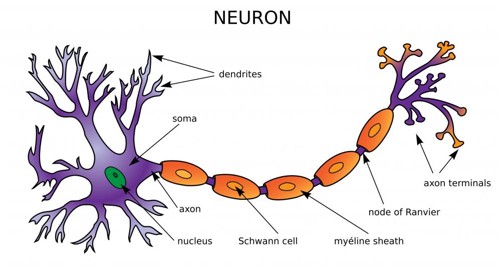
Structure of a motor neuron. Anatomy of a typical human neuron. Structure neuron , SPONSORED,
How do Neurons Work? Neurons lie adjacent to each other but are not connected. There is a tiny gap between neurons called a synapse. The function of a neuron is to transmit nerve impulses along the length of an individual neuron and across the synapse into the next neuron. The electrical signals transmitted by neurons are called action potentials.

Types of Neurones (14.1.2) CIE IGCSE Biology Revision Notes 2023 Save My Exams
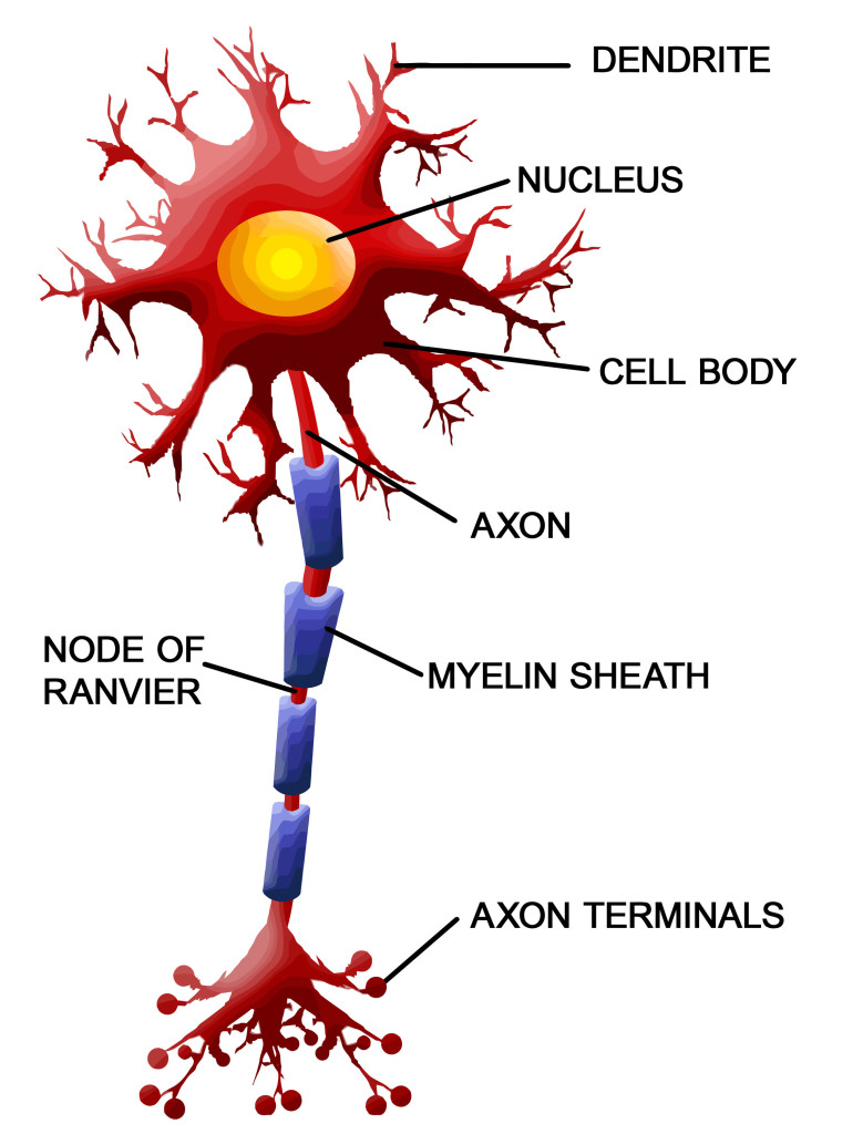
Let's dive a bit deeper into the functioning of motor neurons as we refer to a neatly labeled diagram. Structure, Function, and Location of Motor Neurons Structure All motor neurons are multipolar neurons. A multipolar neuron has only one axon and densely branched dendrites.
:max_bytes(150000):strip_icc()/neuron-anatomy-58530ffe3df78ce2c34a7350.jpg)
Human Biology The Anatomy of Neurons
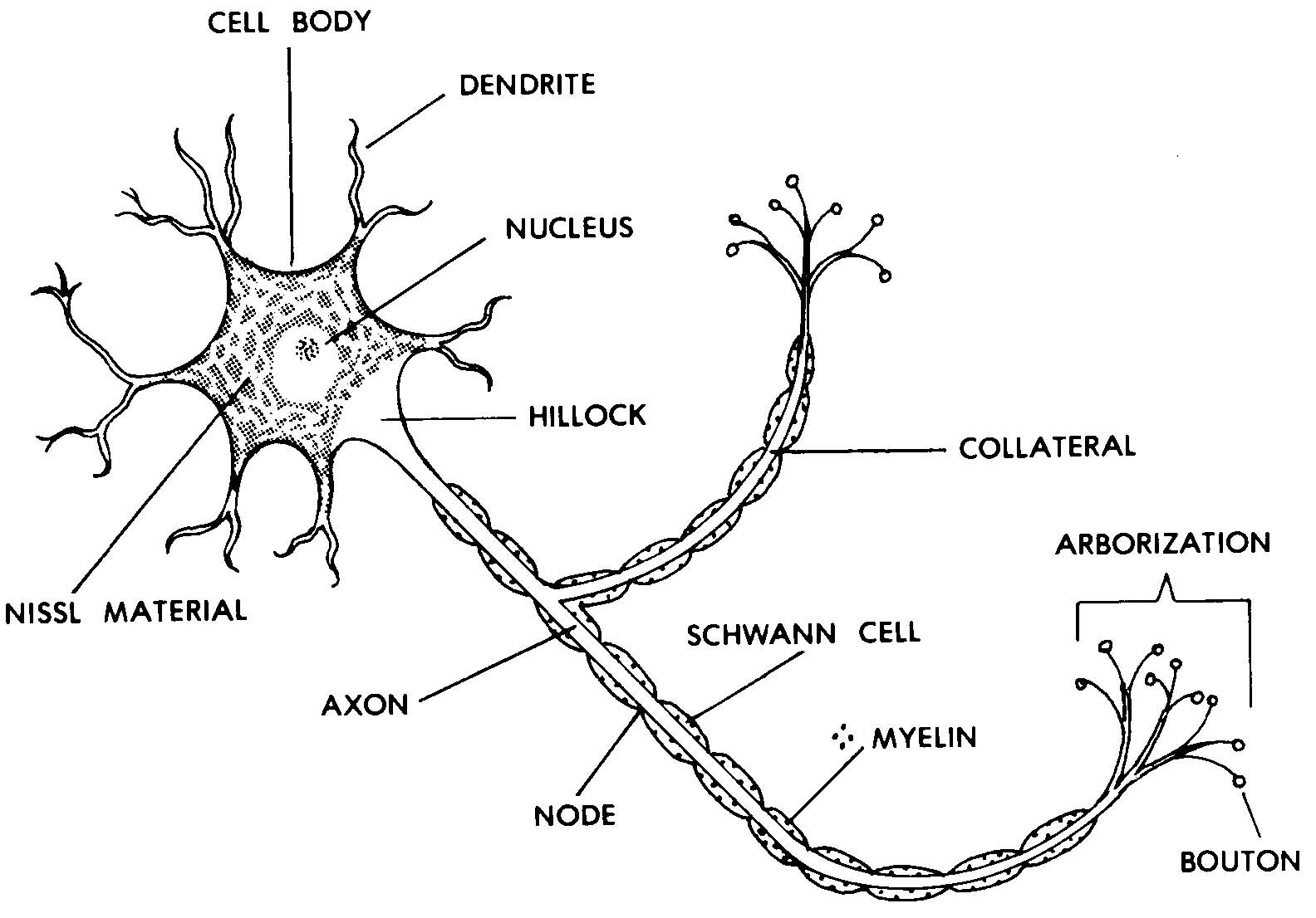
Well-Labelled Diagram of Motor Neuron A motor neuron is a nerve cell that functions to transmit signals from the central area of the nervous system to an effector site such as muscles or glands. A motor neuron can be broadly seen as consisting of three parts - cell body, axon and dendrites.

Neurons What are they and how do they work?
Introduction to neurons and glia. How the structure of a neuron allows it to receive and transmit information. How do you know where you are right now? Your ability to perceive your surroundings - to see, hear, and smell what's around you - depends on your nervous system.
Images 11. Nervous System Basic Human Anatomy
The nervous system is a network of neurons whose main feature is to generate, modulate and transmit information between all the different parts of the human body. This property enables many important functions of the nervous system, such as regulation of vital body functions ( heartbeat, breathing, digestion), sensation and body movements.

Draw A Neuron And Label Its Parts Q10 A Draw The Structure Of Neuron And Label Cell Body And
Cerebellum - molecular, Purkinje, granular layers. Peripheral nerves - epineurium, perineurium, endoneurium. This article will explain the histology of neurons, providing you with information about their structure, types, and clinical relevance. It will also cover briefly the histological layers of the central and peripheral nervous systems.

sensory neurons Google Search Anatomy Pinterest Dental and Dental hygiene
An Easy Guide to Neuron Anatomy with Diagrams Anatomy Types Function Research Takeaway Neurons, also known as nerve cells, send and receive signals from your brain. While neurons have a lot.
The Neuron External Structure and Classification Interactive Biology, with Leslie Samuel
Overall, motor neurons (or motoneurons) comprise various tightly controlled, complex circuits throughout the body that allows for both voluntary and involuntary movements through the innervation of effector muscles and glands. The upper and lower motor neurons form a two-neuron circuit.

Motor Neuron Diagram Neuron Structure and Function_4_ Personal Branding Pinterest
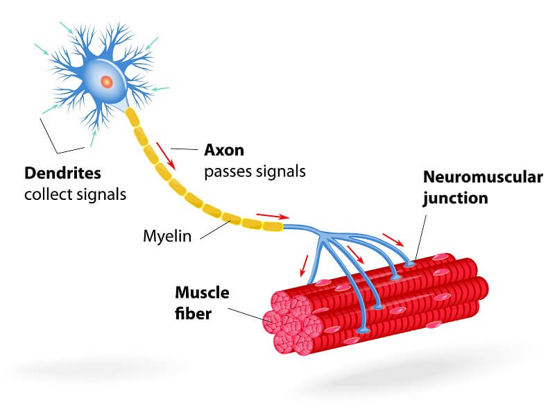
The presence of the synaptic cleft between the synaptic end bulb of the neuron and the motor end plate of the muscle fiber, means that the electrical signal or action potential, arriving from the central nervous system, needs to somehow transverse (cross) this space.The neuromuscular junction accomplishes this by turning the electrical signal from the nervous system into a chemical signal that.

Motor Neuron Disease Neurological Issues Herbal Care Products
motor system: The part of the central nervous system that is involved with movement. It consists of the pyramidal and extrapyramidal systems. cerebral cortex: The gray, folded, outermost layer of the cerebrum that is responsible for higher brain processes such as sensation, voluntary muscle movement, thought, reasoning, and memory.

What Are Dendrites Structure And Its Functions
(A) Diagram showing a lower motor neuron in the spinal cord and the course of its axon to the muscle. (B) Each motor neuron synapses with multiple muscle fibers. The motor neuron and the fibers it contacts defines the motor unit. Cross units and the α motor neurons themselves vary in size.

Figure 7 4 Structure Of A Typical Motor Neuron Bangmuin Image Josh
Summary. Motor neurons are responsible for carrying information from the central nervous system to the peripheral parts of the body. Like other neurons, they have a cell body, an axon, and one or more dendrites. The cell body contains the nucleus of the cell and is the synthetic hub of organelles and macromolecules.
What Are the Major Neurotransmitters? (with pictures)
6.5.2 Draw and label a diagram of the structure of a motor neuron. Figure 6.5.1 - A motor neuron. 6.5.3 State that nerve impulses are conducted from receptors to the CNS by sensory neurons, within the CNS by relay neurons, and from the CNS to effectors by motor neurons.

Neuron Diagram Straight from a Scientist
A motor neuron is a cell of the central nervous system. Motor neurons transmit signals to muscle cells or glands to control their functional output. When these cells are damaged in some way, motor neuron disease can arise. This is characterized by muscle wasting (atrophy) and loss of motor function. Motor Neuron.
Motor Neuron The Definitive Guide Biology Dictionary
A motor neuron (or motoneuron or efferent neuron [1]) is a neuron whose cell body is located in the motor cortex, brainstem or the spinal cord, and whose axon (fiber) projects to the spinal cord or outside of the spinal cord to directly or indirectly control effector organs, mainly muscles and glands. [2] There are two types of motor neuron.

FileNeuron1.jpg Simple English Wikipedia, the free encyclopedia
Diagram Of Neuron with Labels Here is the description of human neuron along with the diagram of the neuron and their parts. The neuron is a specialized and individual cell, which is also known as the nerve cell. A group of neurons forms a nerve.